


When the cells in a person’s bone divide uncontrollably, a mass of invasive tissue, better known as a tumor, develops. While most bone tumors are benign, malignant forms occur most frequently in adolescent/young adult populations. Osteosarcoma is most often sited in the knee or upper arm, and tumors have a high potential to spread, potentially requiring removal of bone segments or full limbs.
3D printing has really helped open up the possibilities for treatment when it comes to bone replacements. Surgery is the most common option for treating bone tumors, and is followed up with reconstruction of bony defects with methods like prosthetic replacement, distraction osteogenesis, intercalary allografting, and fibular grafting. While conventional rigid prosthetics can lead to inflammation, infection, dislocation, and even rejection due to poor surface bioactivity, patient-specific 3D printed prosthetics can offer a much better prognosis.
Recently, a team of medical experts from West China Hospital at Sichuan University published a study, titled “Uncemented three-dimensional-printed prosthetic reconstruction for massive bone defects of the proximal tibia,” in the World Journal of Surgical Oncology about their use of 3D printing to develop a custom knee joint replacement prosthesis for a teenager with metaphyseal osteosarcoma of the tibia.
According to the paper, “Currently, it is challenging to treat massive bone defects of proximal tibia. Although numerous methods are available for reconstruction with epiphysis preservation, limitations in knee function and complications are noted with these methods. Our paper describes our attempt to reconstruct a marked defect in the proximal tibia with an uncemented three-dimensional (3D)-printed prosthesis and to evaluate the prosthesis design and short-term outcomes.
“A 15-year-old boy with metaphyseal osteosarcoma of the tibia underwent intercalary allograft reconstruction following wide tumour resection with epiphysis preservation. However, chronic allograft rejection and/or infection occurred after the surgery and a sinus tract was formed.”
Things turned around for the patient once the initial graft was removed and an antibiotic-loaded cement spacer was implanted, but his limb function still wasn’t up to par.
Due to the odd shape of the defect, and how short the residual proximal tibia was, the team turned to 3D printing to fabricate a custom prosthesis in order to reconstruct the defect and preserve the knee joint. The team learned that a porous structure, with 600μm pore size and 65% porosity, would be able to simulate the real properties of trabecular structures in humans.
- Anteroposterior and lateral radiographs after reconstruction with the 3D printed prosthesis.
- a Anteroposterior radiograph of right tibia 26 months after 3D-printed prosthesis reconstruction. Anteroposterior (b) and lateral (c) T-SMART scanning showed that the 3D prosthesis tightly integrated with the proximal tibia, and partial bone substance could be observed to grow into the porous structure of the prosthesis
Titanium alloys, which offer good mechanical strength, were used at the core of the uncemented prosthesis, in order to improve the matching between the bone defect region and the implant device. To achieve improved bone-impact integration, the stem of the distal part of the 3D printed prosthesis was coated with sophisticated porous bioceramics.
The proximal part was 3D printed on an Arcam Q10 using Electron Beam Melting (EBM) technology, which has seen rising adoption in China and worldwide for prosthetics.
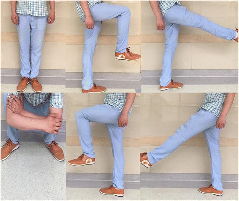
Post-op, the teenager seems to be tolerating the 3D printed prosthetic implant just fine, showing “satisfactory limb function” at his 26-month follow-up appointment and being able to walk, run, and jump with no pain. The prosthesis is fitting well with the real tibia, and there have been so signs of complication – the knee joint is exhibiting motion of 0-130° and an Enneking function score of 93%, which are “comparable” results when compared to normal knee joint motion.
“The 3D-printed prosthesis may be a feasible option in the reconstruction of tibial metaphyseal defects with the preservation of the knee joint. Moreover, it can result in good postoperative function and low complication rates,” the conclusion of the paper reads. “However, a long-term follow-up is required to clarify its long-term outcomes.”
Co-authors of the paper include Minxun Lu, Yongjiang Li, Yi Luo, Wenli Zhang, Yong Zhou, and Chongqi Tu.
Discuss this and other 3D printing topics at 3DPrintBoard.com or share your thoughts below.
[Source: Medical News Bulletin / Images: West China Hospital, Sichuan University]
When it comes to 3D real estate visualization in the USA, our service provides the perfect solution for bringing property listings to life. Through our platform, you can easily access cutting-edge 3D renderings that showcase your real estate projects in a way that attracts potential buyers and investors. Whether it's residential, commercial, or mixed-use properties, our team of experts uses advanced technology to create immersive visualizations that highlight the best features of your property, making it easier for clients to imagine the space as their own.
Through our website, you can quickly get high-quality 3D real estate visualizations that are tailored to your specific needs. With our help, you'll stand out in the competitive real estate market by offering potential buyers a realistic, interactive view of your property. Our efficient process ensures a fast turnaround time, while our attention to detail guarantees that every aspect of the property is represented accurately, giving you a powerful marketing tool to promote your real estate listings.


